
4 Microscopic Exam of Urine (Part 1/2) YouTube
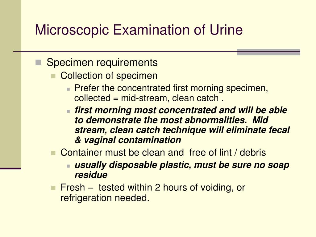
Microscopic Urinalysis Does this test have other names? Microscopic urine analysis, microscopic examination of urine. What is this test? This test looks at a sample of your urine under a microscope. It can see cells from your urinary tract, blood cells, crystals, bacteria, parasites, and cells from tumors.
Urine Sediment of the Month Renal Fellow Network
In Medical Laboratory Science (MLS) Urinalysis course, students learn to evaluate normal and abnormal formed urinary elements through microscopic examination of urine sediment. They learn to interpret results and correlate with other laboratory data to identify disease. Access to quality atlases are a vital tool in the students' learning process.
PPT Microscopic Examination of Urine PowerPoint Presentation, free download ID236160
A urinalysis is a test of your urine. It's used to detect and manage a wide range of disorders, such as urinary tract infections, kidney disease and diabetes. A urinalysis involves checking the appearance, concentration and content of urine. For example, a urinary tract infection can make urine look cloudy instead of clear.
Urine Sediment of the Month Findings in Cirrhosis, Cholestasis, and Hyperbilirubinuria Renal
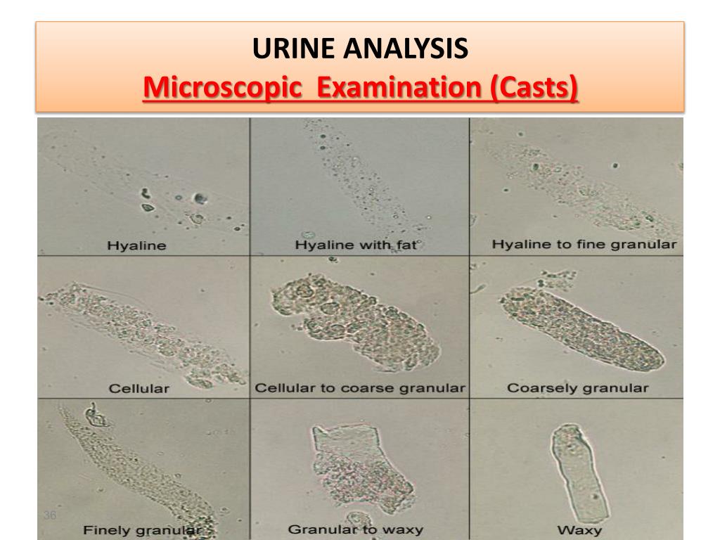
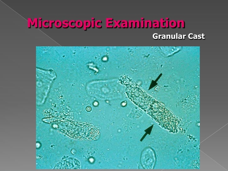
0:00 / 7:58 An introduction to the microscopic component of the UA, which looks at the cells (WBC, RBC, epithelia, bacteria, and yeast) and casts (hyaline, cellular and.
Approach to the Patient With Renal Disease Part 2
This course covers the basics of urine microscopic examination, including numerous brightfield and phase-contrast images of urinary sediment elements. It is assumed that students have a basic knowledge of urinalysis macroscopic and dipstick examination. The course covers specimen collections and processing, casts, cellular elements, normal and.
PPT Urinalysis PowerPoint Presentation, free download ID2773692
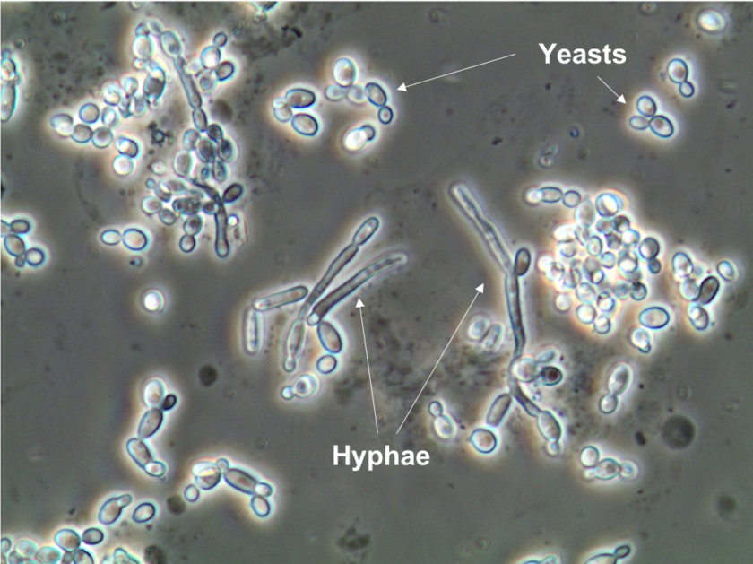
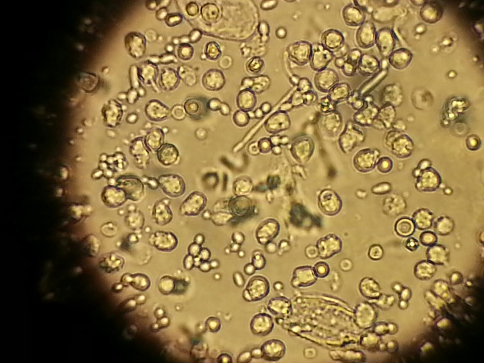
Browse 1,100+ urine microscopy stock photos and images available, or start a new search to explore more stock photos and images. Sort by: Most popular Microscopic image showing Calcium oxalate (monohydrate and. Pseudohyphae and budding yeast cells in urine
PPT Urine analysis PowerPoint Presentation ID632788
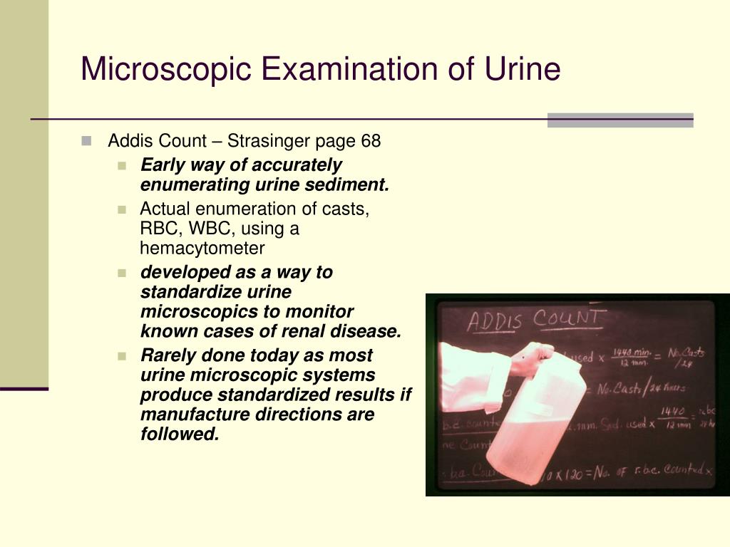
The standardized quantitative microscopic examination of urine sediment made its clinical laboratory debut in 1926. At that time, Thomas Addis developed a procedure to quantify formed elements in a 12-hour overnight urine collection. The purpose of this test, the Addis count, was to follow the progress of renal diseases, particularly acute.
Urinalysis Principle ,Procedure Result interpretation Including Microscope
Urine AnalysisSample Collection and Microscopic Examination. Urine analysis is the term used to refer to the test used to evaluate a urine sample. Typically, this test is used for the purposes of assessing a wide range of disorders, which may include kidney disease, urinary tract infection (UTI) dehydration as well as diabetes.
Hyphae in routine urine microscopic examination medlabprofessionals
The urine microscopic exam is difficult to teach because supervised instruction and textbook-based teaching suffer from numerous drawbacks. Here, we describe Urinalysis-Tutor, a computer program that uses digitized microscope images and computer-based teaching techniques to systematically teach the urine microscopic exam.

Understanding Urine Microscopic Examination Importance and Interpretation
Urine microscopic examination is a laboratory test that analyzes a small sample of urine under a microscope to evaluate the physical, chemical, and microscopic characteristics of the urine. It helps to diagnose various health conditions, such as urinary tract infections, kidney disease, and bladder cancer.
PPT Microscopic Examination of Urine PowerPoint Presentation ID236160
Microscopic exam: With these tests, a small sample of urine is examined under a microscope for abnormal crystals, bacteria, or cell types. Infections and kidney problems are the most common.

Urinalysis Microscopic Examination All Things Kidney Official
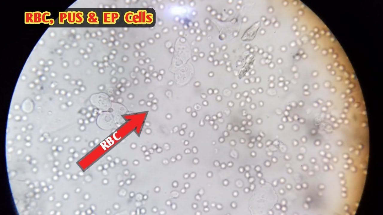
177 urine microscopic examination stock photos, 3D objects, vectors, and illustrations are available royalty-free. See urine microscopic examination stock video clips Filters All images Photos Vectors Illustrations 3D Objects Sort by Popular Urine microscopic examination 40x show plenty of pus cells and few epithelial cells. Micrograph
Pus Cells In Urine Microscopy
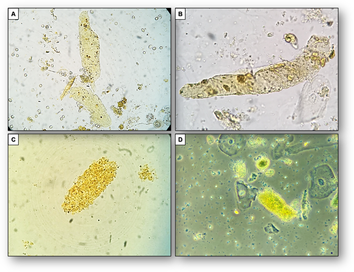
Figure 1. Photomicrographs of Urine Sediments Obtained with the Use of Digital Cameras and Cell-Phone Cameras. We recently determined that we could photograph the urine sediment through a.

Cells In Urine Sediment / Microscopic examination of urine sediment ©this article describes
Check out Dr. Jay Seltzer's KIDNEYcon 2022 Urine Microscopy video here with dazzling images! Images are organized in the sections below: Unstained Casts & Acanthocytes Stained Casts & Acanthocytes Phase Contrast Casts & Acanthocytes Squamous, Transitional, and Renal Tubular Epithelial Cells Lipids Crystals Other
Examination Pus Cells In Urine Microscopy
Urine microscopy is an important tool for the diagnosis and management of several conditions affecting the kidneys and urinary tract. In this review, we describe the automated instruments, based either on flow cytometry or digitized microscopy, that are currently in use in large clinical laboratories. These tools allow the examination of large numbers of samples in short periods.
The Microscopic Examination for urine sediment Medical Labs Everyday
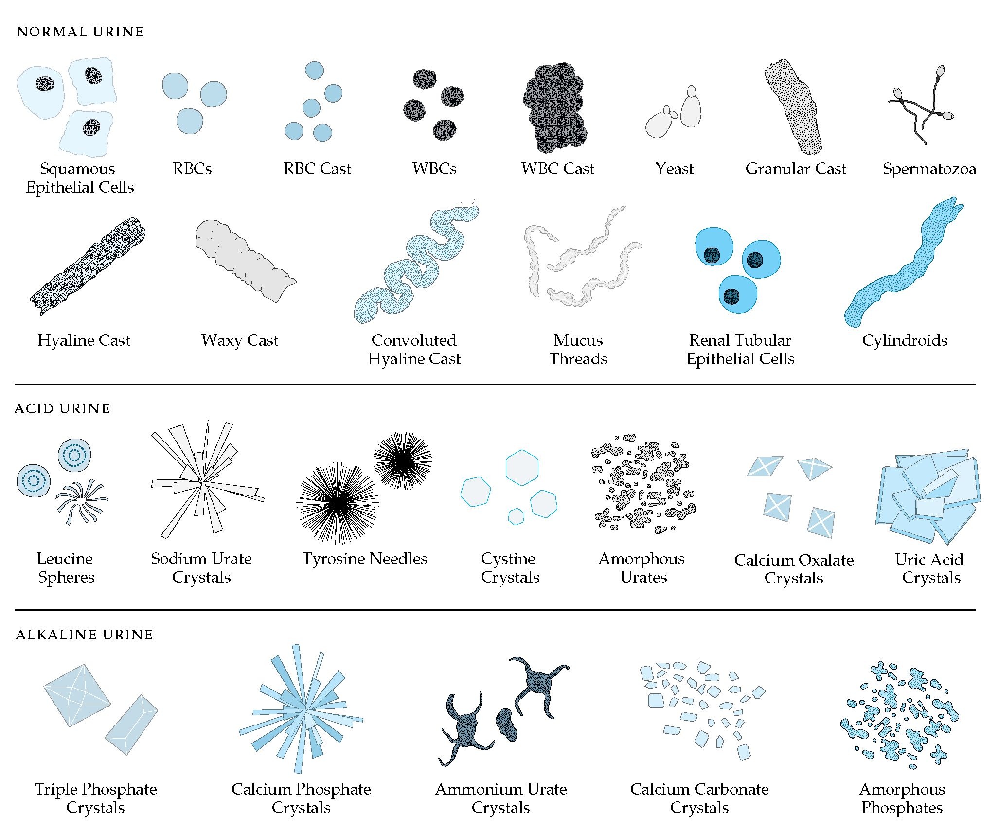
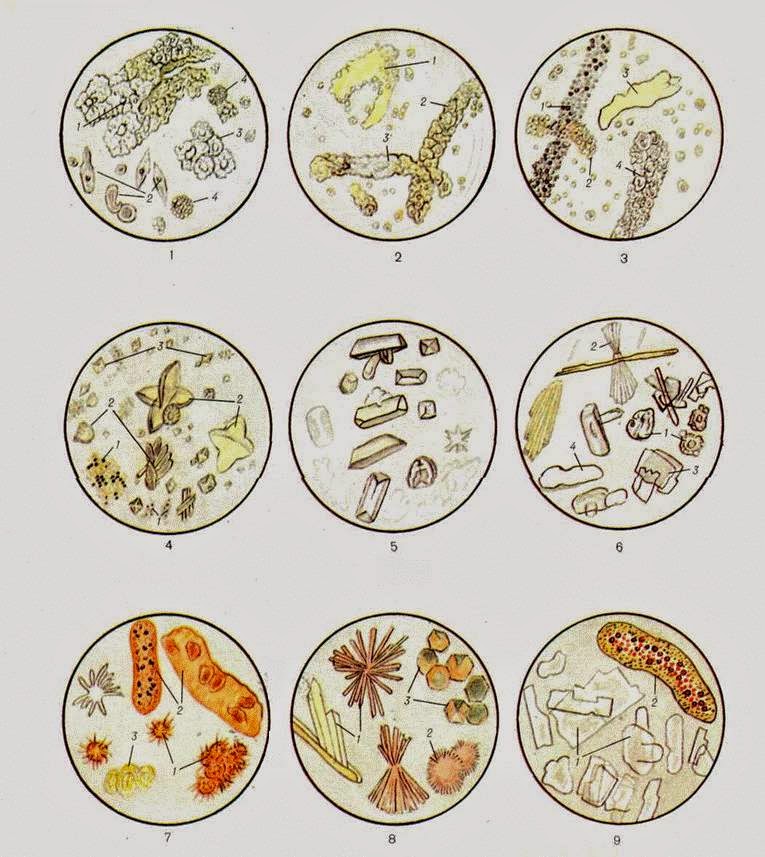
Examination of microscopic urine sediment images resolve various constituents present in the urine such as Red blood cells (RBC), White blood cells (WBC) or Leukocytes, epithelial cells crystals, and casts. The findings from microscopic examination is used to diagnose several renal diseases. 2. Manual microscopic examination